
Microscopic Lung Cell Footage Earns Top Honor in Nikon’s 2026 Motion Competition
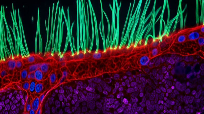
Qwenews.com – A close-up look at the hidden mechanics of the human airway has taken the top prize in the 16th annual Nikon Small World in Motion competition. The winning video reveals the irregular movement of microscopic cilia in a child with primary ciliary dyskinesia, a rare inherited condition that can interfere with the lungs’ ability to clear mucus and other material from the airways.
The footage was created by Ning Xu of Tsinghua University in Beijing, China. Xu used an advanced microscopy technique to capture the activity of cilia, the tiny hairlike projections found on the surface of certain cells throughout the body. In healthy airways, these structures move in organized, rhythmic patterns that help push mucus and trapped debris outward.
For people with primary ciliary dyskinesia, often called PCD, that coordinated motion may be disrupted. Cilia can move in an unusual way or fail to beat effectively, making airway clearance more difficult. The result can be recurrent respiratory infections, as mucus and particles are not removed as efficiently from the lungs.
Why Motion Matters in Microscopy
The winning entry demonstrates why a moving image can offer insights that a still frame cannot. The cilia themselves are far too small to observe with the unaided eye, but their behavior becomes immediately visible when magnified and recorded over time. Their changing motion helps illustrate the difference between a coordinated biological process and one affected by disease.
“To understand this disease, you need to observe around 10 seconds of motion, not just a still image,” Xu, an optical engineer who develops advanced imaging systems to aid scientists in better understanding complex cellular structures through motion, said in a press release Wednesday.
Microscopy is often associated with static images of cells, tissues and microorganisms. Yet many essential processes inside living organisms depend on movement: cells divide, organelles shift, fluids circulate and structures such as cilia beat in repeating cycles. Video microscopy can make those processes easier for researchers, students and the public to recognize.
“When people watch cilia beating, they immediately understand that something beautiful and important is happening inside us,” Xu added. “I think this competition turns microscopy into a shared language.”
That combination of scientific value and visual appeal sits at the center of Nikon Small World in Motion. The competition was established to celebrate advances in microscopic photography and video, while also inviting participants with an interest in imaging to submit their work. Its entries frequently show phenomena that are normally inaccessible without specialized equipment, transforming tiny biological events into striking visual stories.
A Global Field of Scientific Images
This year’s video competition drew 347 submissions from 40 countries. A judging panel of experts in science and advanced light microscopy selected the winners. The range of entries reflected the breadth of activity visible at microscopic scales, from the behavior of individual cells to the movement of small organisms and plant structures.
Eric Flem, senior manager of communications and CRM at Nikon Instruments, said the winning work reflects the long-running purpose of the contest: to spark curiosity, support scientific exploration and highlight the creative side of research.
“Xu’s work, and the work of all our winners, reflects the competition’s enduring purpose to inspire wonder, fuel discovery, and showcase the artistry inherent in scientific exploration,” Flem said in the release.
Xu’s video places a medical condition in a highly visual context. Rather than presenting PCD only through clinical terminology, it lets viewers see a cellular mechanism that is central to airway health. The images do not replace diagnosis or treatment, but they offer a clear illustration of why normal ciliary function matters.
Cilia perform important work in the respiratory system. Their coordinated beating creates a transport process along airway surfaces, helping move mucus away from the lungs. Mucus can trap dust, microbes and other particles inhaled during breathing. When that clearing system does not function properly, the airway can become more vulnerable to repeated infection and related complications.
Other Prize-Winning Views of Life at Small Scale
Second place went to Nguyen Nam Nhat, whose entry featured a tiny roundworm alongside a single-celled Dileptus. The pairing offered another example of the activity that can unfold beyond ordinary human vision, with organisms moving and interacting at a scale that requires magnification to appreciate.
Benedikt Pleyer received third place for a video of jellyfish larvae held within water droplets. The entry highlighted the delicate appearance of early marine life and the unusual visual effects created when living material is suspended in small amounts of water.
Andrew Moore was awarded fourth place for recording synchronized division among neighboring cells. Cell division is one of biology’s most fundamental processes, and the synchronized event captured in the video showed how timing and organization can emerge within closely situated cells.
Fifth place was awarded to Patrick Hickey for a video showing mitochondria in green and chloroplasts in red inside turtle vine leaf cells. Mitochondria are associated with cellular energy production, while chloroplasts are the structures that enable plants to capture light energy for photosynthesis. The contrasting colors made their movement and arrangement easier to follow.
Together, the selected videos show how microscopy can bridge research and public understanding. A microscopic view may begin as specialized scientific work, but motion, color and magnification can make it legible to viewers without a laboratory background. In Xu’s winning footage, an airway disorder becomes not merely an abstract diagnosis, but a visible change in the finely coordinated movement taking place inside the body.
Related Reading
Frequently Asked Questions
What is Astonishing footage of abnormal cell movement?
Astonishing footage of abnormal cell movement is the main topic of this guide. The article explains the context, practical details, and next steps readers should understand.
Why does Astonishing footage of abnormal cell movement matter?
Astonishing footage of abnormal cell movement matters because readers are looking for a useful answer, not just a short summary. Good content should match search intent and help them decide what to do next.